In a recent patch test — a routine procedure to determine which substances could be triggering skin flare-ups — just a few faint pink and purple hues showed up. To the casual eye, the 47-year-old African American patient had “very mild erythema,” said Ama Alexis, MD, an allergist-immunologist at NYU Langone Health in New York City. Yet during that November office visit, the patient said she was so itchy that she was “barely able to sleep.”
Some 16.5 million people in the United States suffer from atopic dermatitis. The disease affects 15%-20% of children and 1%-3% of adults globally, and rates have risen two- to threefold in industrialized nations since the 1970s.
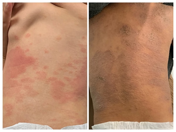
Erythema on a 10-month-old African American male (right) with atopic dermatitis has darker hues compared with the bright red discoloration on an age-matched white infant (left). Using redness to assess erythema can lead investigators to underestimate disease severity in research trials.
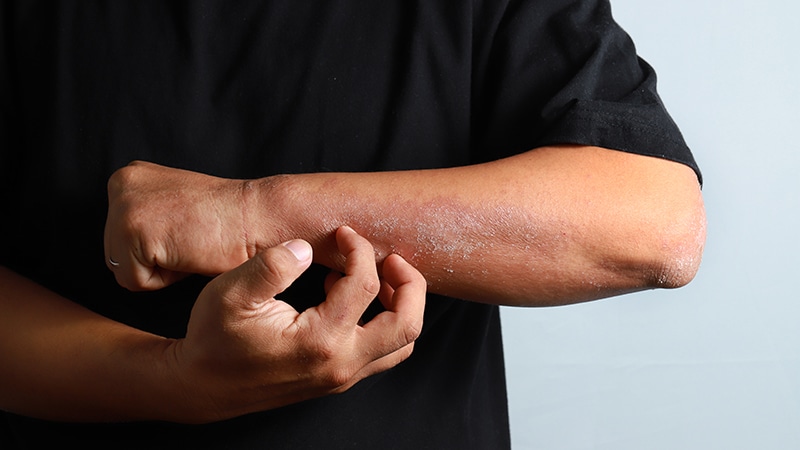
Doctors are classically trained to recognize atopic dermatitis as red blotches on the skin. However, in patients with darker complexions, rashes may not look entirely red or not red at all. Often the erythema will appear violet or gray or dark brown. “With that lack of bright red, sometimes it’s hard to assess the acuity of the lesions because it just doesn’t look as alarming,” said Alexis, who is also a clinical assistant professor of pediatrics at NYU Grossman School of Medicine. “But it may feel alarming.”
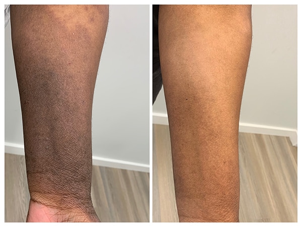
Left image: Maroon, grey and purple hues — rather than bright red — are common in atopic dermatitis patients with skin of color. Right image shows baseline skin color after several weeks of treatment.
Those instances of disconnect between how a patient feels and what a physician sees can create challenges in both clinical and research settings. In the clinic, doctors who aren’t used to recognizing erythema across a range of skin tones could underestimate the seriousness of a patient’s condition, which can lead to delayed diagnoses or inappropriate treatment. And in research studies, which use erythema as a key component of patient outcome metrics, it’s also possible to underestimate disease severity — and that can skew trial results that determine which drugs get approved.
“It takes some adjustment and training of the eye to be able to detect the nuances of what erythema looks like across the whole spectrum of different pigmentations,” said Andrew Alexis, MD, MPH, professor of clinical dermatology at Weill Cornell Medical College in New York City. He is married to Ama Alexis and spoke about atopic dermatitis in skin of color at the 3rd Annual Revolutionizing Atopic Dermatitis (RAD) Virtual Conference in December.

Dr Andrew Alexis
Skin disease images that are found online or in medical textbooks mostly show white patients, yet atopic dermatitis can be more common and more severe in certain other ethnic groups. In multiple studies in the United States and United Kingdom, Black children are about twice as likely to develop the skin disorder than their White counterparts. In addition, Black and Hispanic children in the US are more likely than White children to visit a primary care doctor or emergency room for atopic dermatitis. And among adults, Blacks are three times more likely and Asian/Pacific Islanders nearly seven times more likely than White people to see a physician for this condition.
Reasons for these disparities are complicated and still being worked out.
Beyond Genetics
Genetic ancestry does not seem to explain the higher rates of atopic dermatitis in African Americans. That was the upshot of a 2020 analysis of data from two multiethnic cohorts — 86,893 adults in a large Kaiser Permanente study not specific to eczema (GERA: Genetic Epidemiology Research on Adult Health and Aging), and 5467 subjects aged 2-26 years from the national Pediatric Eczema Elective Registry (PEER).

Dr Katrina Abuabara
Controlling for household income in the pediatric cohort, Black patients were more likely to have eczema and their disease was more severe and more poorly controlled than White counterparts. And in the Kaiser GERA cohort, after controlling for income and education levels, “you had about twice the risk of having atopic dermatitis if you self-identified as African American or Black, versus White,” said study leader Katrina Abuabara, MD, MA, MSCE, associate professor of dermatology at UCSF and associate adjunct professor of epidemiology at UC Berkeley School of Public Health.
African American individuals show considerable range in “how African or how European” their genomes are, yet when atopic dermatitis diagnoses were analyzed against this continuous measure of genetic ancestry, “it was not at all predictive,” Abuabara told Medscape Medical News.
Even after zeroing in on single-nucleotide polymorphisms that relate to skin color or specific variants that associated with higher atopic dermatitis risk in prior studies, none of those genetic markers could explain the disparities. “It was only the self-identified race or ethnicity, which obviously encompasses more than just genetics,” said Abuabara.
Although genetic ancestry may not account for the higher burden of atopic dermatitis in some racial/ethnic groups, some research does suggest that Black skin has distinct molecular and clinical features. In a study published last May, researchers at Johns Hopkins University in Baltimore collected skin biopsies from patients with healthy skin or moderate to severe atopic dermatitis.

Dr Shawn Kwatra
From a slew of histological and gene expression analyses, they found that African American patients with atopic dermatitis had higher levels of ferritin, blood eosinophils, and C-reactive protein compared with White patients with atopic dermatitis and African Americans with healthy skin. These characteristics point to “higher systemic inflammation” in African American patients, said study leader Shawn Kwatra, associate professor of dermatology and oncology at Johns Hopkins.
In Black patients, atopic dermatitis can also look quite different from hallmark features described in diagnostic criteria based on predominantly White populations. For example, Hanifin & Rajka and UK Working Party criteria specify involvement of flexural surfaces — skin on the side of joints that bend, such as inside the elbow and behind the knee. In African Americans, though, atopic dermatitis often affects extensoral surfaces — the front of the knee and backs of hands and elbows.
Black patients with atopic dermatitis commonly develop prurigo nodularis — itchy bumps that are not in typical diagnostic criteria.
Plus, Black patients commonly present with prurigo nodularis — yet these itchy bumps are not in the diagnostic criteria, said Kwatra, who also directs the Johns Hopkins Itch Center and co-authored a 2021 review on pruritis (severe itch) in Black skin. “These are major groups with major guidelines that are based almost entirely on the presentation of atopic dermatitis in Caucasians.”
Tools Fall Short
It’s no surprise, then, that nuanced disease presentations in skin of color can go underrecognized. This happened in a clinical phenotyping study involving 592 adults diagnosed with atopic dermatitis. Participants completed questionnaires about itch severity, skin symptoms, and quality of life, then got examined by a dermatologist who assessed their disease severity using validated tools such as the investigator global assessment (IGA) scale, eczema area and severity index (EASI), and the SCORing Atopic Dermatitis (SCORAD) scale.
Based on these patient-reported outcomes and physician assessments, researchers grouped participants into four subsets: mild-moderate itch and lesions (MI/ML), mild-moderate itch and severe lesions (MI/SL), severe itch and mild-moderate lesions (SI/ML), and severe itch and severe lesions (SI/SL). Looking back at self-identified race and ethnicity in these patients’ records, the team discovered an overrepresentation of Black patients in the SI/ML group.

Dr Jonathan Silverberg
These patients are “reporting that they have severe itch, but I can’t see it,” said study leader Jonathan Silverberg, MD, PhD, MPH, associate professor of dermatology at George Washington University in Washington, DC. The study shows that “the outcome measures are just missing things, missing important subsets of patients,” Silverberg told Medscape Medical News. “If we rely exclusively on these lesional outcomes to tell us how well a drug is working, we may be missing the boat.”
As a first step to seeing how assessment tools perform in patients of color, Silverberg teamed up with Andrew Alexis and others to comb through the literature assessing the data on widely used atopic dermatitis outcome measures across race, ethnicity, and skin tone. A key takeaway: “There wasn’t a single measure that actually had really rich data across diverse patient populations,” Silverberg said. “And if they did, they didn’t even document it. This is such a problem. It’s not even on investigators’ radars.” Those findings were presented last summer at the Revolutionizing Atopic Dermatitis conference, and later published in the Journal of Investigative Dermatology.
Not only are Black patients developing atopic dermatitis more often, suffering more from the condition, and being inappropriately assessed — they’re also not benefiting as much from newly available medications. In an analysis of medical records of patients who were prescribed treatments for various skin conditions, Kwatra and colleagues found that Black patients were less likely to receive dupilumab (Dupixent), a frontline therapy approved in 2017 to treat moderate to severe atopic dermatitis. In addition, they were less likely to get several topical medications (crisaborole [Eucrisa], pimecrolimus [Elidel], tacrolimus [multiple brands]) but instead were prescribed hydrocortisone, the mildest treatment, more than twice as often as White patients.

Dr Ama Alexis
Considering the earlier prevalence studies along with this recent analysis of therapies, “there’s a disconnect between the disease state that we’re seeing and the medication that we’re giving,” said Ama Alexis.
One of the big challenges is that itch — one of the most burdensome aspects of atopic dermatitis — is a complex symptom that is hard to measure. Some patients are intensely itchy for 30 minutes a day where others have milder, nagging itch 24 hours a day, and still others may have “10 out of 10 itch or pain for 24 hours a day,” Silverberg said. “We have to recognize that when we use these kinds of very crude assessments, we’re not getting the whole story.”
Harmonizing Outcome Measures for Eczema (HOME) — an international group that comes up with consensus tools to assess outcomes in clinical trials — does recommend several patient-reported measures, including one for itch intensity. Patients “know their lived experience. They know how bad their disease is,” Silverberg said. “Patient-reported outcomes definitely need greater prioritization and attention in this disease.”
However, the primary endpoints for FDA and EMA trials “are virtually always measures of signs (IGA and EASI),” said Silverberg, whose group has validated several itch markers, including one called SCORAD-scratch , that have yet to gain traction with regulatory bodies.
More Education Needed
One approach to addressing the disconnect between patient- and physician-reported outcomes “may be to simply better educate clinicians and investigators on how to use the existing tools to more accurately assess their patients,” said Andrew Alexis, who is vice chair for diversity and inclusion for the department of dermatology at Weill Cornell Medicine.
Toward this end, he and Silverberg have created an Eczema Area and Severity Index (EASI) lesional severity atlas with representative images showing clinical signs of atopic dermatitis across a range of skin tones. They presented the atlas on a poster at the Revolutionizing Atopic Dermatitis (RAD) Virtual Conference last December, and have a paper under review.
Their efforts add to a growing collection of resources to help reduce health disparities in people of color. Numerous texts, including a handbook called Mind the Gap created by a Black medical student, are available to assist with diagnoses in skin of color. The medical software company VisualDx has launched a global initiative, Project IMPACT, to collect images from diverse populations.
“Everybody would benefit from more education,” said Ama Alexis. Education would help doctors “be able to easily recognize your condition regardless of your skin color” and “for patients, to not think their disease is so unique that they can’t even find it when they google it.”
As for the aforementioned 47-year-old Black woman who received patch testing last fall, her skin was positive in four locations. With side lighting, Alexis said she could see and palpate faint purple papules in the square that corresponded with several fragrances (hydroperoxides of limonene and hydroperoxides of linalool). This patient had recently started using a hair conditioner containing these ingredients. Once she stopped using the conditioner, her eczema improved.
Resources:
Skin of Color dermatology textbooks
Eczema in Skin of Color (American College of Allergy, Asthma & Immunology; Allergy & Asthma Network)
Training, articles, images (British Association of Dermatologists)
VisualDx (software with medical images and skin of color atlas to help diagnose conditions in diverse patients)
Skin Deep (open-access photos of medical conditions in a range of skin tones)
Abuabara receives consulting fees from Target RWE, and her institution receives grant funding from Pfizer and LaRoche Posay.
Ama Alexis reports financial relationships with Regeneron and Pfizer.
Andrew Alexis has received grants (funds to institution) from Leo, Novartis, Almirall, Bristol-Myers-Squibb, Amgen, Menlo, Galderma, Valeant (Bausch Health), Cara, Arcutis, and Dermavant. He has consulted or served on advisory boards for Leo, Galderma, Pfizer, Sanofi-Regeneron, Dermavant, Beiersdorf, Valeant, L’Oreal, BMS, Bausch Health, UCB, Vyne , Arcutis, Janssen, Allergan, Almirall, AbbVie, Sol-Gel , Amgen, VisualDx, Eli Lilly; and received speaking fees from Regeneron, Sanofi-Genzyme, and Pfizer.
Kwatra is a member of the Board of Directors of the Skin of Color Society and serves as an advisory board member/consultant for AbbVie, Celldex Therapeutics, Galderma, Incyte Corporation, Johnson & Johnson, Novartis Pharmaceuticals Corporation, Pfizer, Regeneron Pharmaceuticals, Sanofi, and Kiniksa Pharmaceuticals and has served as an investigator for Galderma, Pfizer, and Sanofi.
Silverberg has received honoraria as a consultant and/or advisory board member for AbbVie, Afyx, Aobiome, Arena, Asana, Aslan, BioMX, Bluefin, Bodewell, Boehringer Ingelheim, Celgene, Connect Biopharma, Dermavant, Dermira, Eli Lilly, Galderma, GlaxoSmithKline, Incyte, Kiniksa, Leo Pharma, Luna, Menlo, Novartis, Pfizer, RAPT, Regeneron, Sanofi-Genzyme; has been a speaker for AbbVie, Eli Lilly, Leo Pharma, Pfizer, Regeneron, Sanofi-Genzyme; and has received grants from Galderma, Pfizer.
Esther Landhuis is a freelance science journalist in the San Francisco Bay Area. She can be found on Twitter @elandhuis.
For more news, follow Medscape on Facebook, Twitter, Instagram, YouTube, and LinkedIn
Source: Read Full Article