
Each distinct part of the brain plays a different role in allowing humans to have thoughts and memories, move their arms and legs, sense smell, sight, hearing, touch, and taste, as well as maintain the functions of many organs within the body.
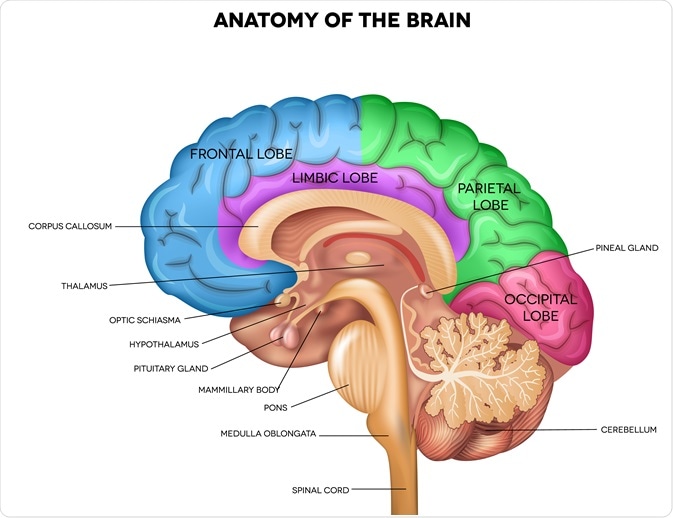
Image Credit: Tefi/Shutterstock.com
The cells of the brain
The two predominant cell types that the brain is comprised of include neurons and glial cells, the latter of which can also be referred to as neuroglia or glia. Depending upon their location throughout both the central and peripheral nervous systems (CNS and PNS, respectively), neurons can have varying morphologies.
While this may be true, all neurons will share four common regions that include the cell body, dendrites, axon, and axon terminal, each of which has their own respective functions. The cell body of the neuron, for example, has a nucleus that is responsible for the synthesis of proteins that travel through microtubules down to the axons and axon terminals through a process known as anterograde transport.
Glial cells can be further subdivided into oligodendrocytes, microglial cells, and astrocytes, the latter of which is the most abundant type of glial cell and occupies approximately 25% of the total brain volume. Astrocytes can be further classified as either protoplasmic or fibrous. Protoplasmic astrocytes are present in the gray matter of the brain and have several branches that can interact with both synapses and blood vessels.
Comparatively, fibrous astrocytes, which can only be found in the white matter of the brain, have long fiber-like processes that also interact with blood vessels, as well as the nodes of Ranvier. Taken together, both types of astrocytes interact with blood vessels by adjusting their blood flow in response to synaptic activity.
Like the Schwan cells of the PNS, oligodendrocytes, which are only present in the CNS, are responsible for the production of myelin, which maintains the electrical impulse conduction of nerve signals and maximizes their velocity as needed.
The final type of glial cells includes microglia, which is part of the macrophage population of the CNS that also includes perivascular macrophages, meningeal macrophages, macrophages of the circumventricular organs (CVO), and the microglia of the choroid plexus. Like any other type of macrophage, microglia are immune phagocytic cells and thus function to protect the CNS from potential pathogens.
Divisions of the brain
The human brain can often be divided into three distinct parts, which include the cerebrum, the cerebellum, and the brainstem.
The cerebrum
The major portion of the brain is the cerebrum, which divides the left and right cerebral hemispheres, both of which have numerous folds and convolutions present on their surface. Between these convolutions are ridges known as gyri. Small grooves that are present between the gyri are known as the plural of sulcus or sulci, whereas larger grooves are referred to as fissures.
The right and left cerebral hemispheres, both of which are covered in the cerebral cortex that is otherwise known as gray matter, are joined together by the corpus callosum. Whereas the left hemisphere controls speech and abstract thinking, the right hemisphere controls spatial thinking.
The frontal, parietal, temporal, and occipital lobes are the four lobes that make up the cerebrum. The frontal lobes, which are present directly behind the forehead, are the largest lobes of the human brain. The frontal lobes are primarily responsible for controlling language, motor function, and various cognitive processes including self-awareness, mood, affect, memory, attention, as well as both social and moral reasoning.
Within the frontal lobe is Broca’s area, which is responsible for speech production. The parietal lobes, who can be found near the center of the brain between the frontal and occipital lobes, are responsible for interpreting different sensory and memory functions.
The temporal lobes, which is commonly referred to as the neocortex, is located close to the base of the skull. Within the temporal lobe is the Wernicke area, which allows individuals to understand both spoken and written language. In addition to processing speech, the temporal lobe also processes sensory information that contributes to the retention of memories, languages, and emotions.
The fourth and final lobe of the cerebrum is the occipital lobe, which is the smallest lobe of the cerebrum and forms the caudal part of the brain. The primary function of the occipital lobe is the interpretation of visual information.
The cerebellum
The largest of the hindbrain is the cerebellum. Upon reception of motor information from both the cerebral cortex and the musculoskeletal structures of the body, the cerebellum coordinates these signals to maintain the gait and posture of humans in motion.
Although the cerebellum itself does not initiate muscle contraction, it aids in the refinement and accuracy of motor activity by controlling muscle tone. In addition to its role in controlling balance and regulating motor movement, the cerebellum also plays a role in the regulation of fear and other cognitive functions such as attention, language, and the human response to pleasure.
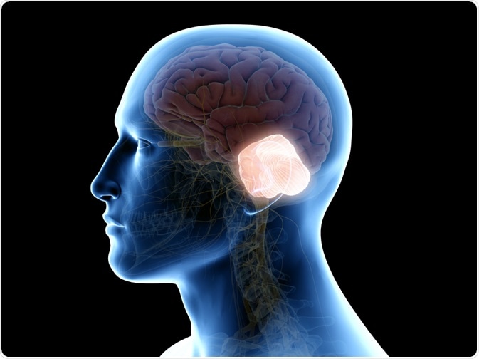
Image Credit: SciePro/Shutterstock.com
The brainstem
The cerebellum and spinal cord are connected to the cerebral hemispheres by the brainstem. The brain stem can be classified into four distinct sections that include the diencephalon, midbrain, pons, and medulla oblongata. The diencephalon, which is the most superior portion of the brainstem, is further subdivided into four portions that include the epithalamus, subthalamus, hypothalamus, and thalamus.
The thalamus, which is the largest portion of the diencephalon, serves as a relay point for all sensory information that enters the cortex and eventually gets transmitted to the cerebrum for processing. The hypothalamus also processes incoming sensory information; however, all of the information processed by the hypothalamus is derived from the autonomic nervous system (ANS).
As a result, the hypothalamus maintains eating habits, sexual behavior, and sleep patterns in addition to maintaining an individual’s body temperature. Additionally, the secretions of the pituitary gland, which develops from a downward extension of the hypothalamus, is controlled by the hypothalamus.
The midbrain, which connects the diencephalon to the pons, controls ocular motion, whereas the pons is involved in the regulation of eye and facial movements, hearing and balance, as well as all sensory information processed by the facial nerves.
The medulla oblongata, which is located between the pons and the spinal cord and is therefore the most inferior portion of the brainstem, controls autonomic functions such as breathing, blood pressure, cardiac rhythms, and swallowing. Notably, brain death of patients in a clinical setting is declared when there is significant destruction of the medulla oblongata.
References and Further Reading
- Anatomy of the Brain. [Online]. Available from: https://www.aans.org/en/Patients/Neurosurgical-Conditions-and-Treatments/Anatomy-of-the-Brain.
- Maldonado, K. A., Alsayouri, K. (2020). Physiology, Brain. In: StatPearls. Available from: https://www.ncbi.nlm.nih.gov/books/NBK551718/.
- Jimsheleishvili, S., & Dididze, M. (2020). Neuroanatomy, Cerebellum. In: StatPearls. Available from: https://www.ncbi.nlm.nih.gov/books/NBK538167/.
- Messé, A., Rudrauf, D., Benali, H., Marrelec, G. (2014). Relating structure and function in the human brain: relative contributions of anatomy, stationary dynamics, and non-stationarities. PLoS Computational Biology 10(3):e1003530. doi:10.1371/journal.pcbi.1003530.
Last Updated: Dec 20, 2020

Written by
Benedette Cuffari
After completing her Bachelor of Science in Toxicology with two minors in Spanish and Chemistry in 2016, Benedette continued her studies to complete her Master of Science in Toxicology in May of 2018.During graduate school, Benedette investigated the dermatotoxicity of mechlorethamine and bendamustine, which are two nitrogen mustard alkylating agents that are currently used in anticancer therapy.
Source: Read Full Article


