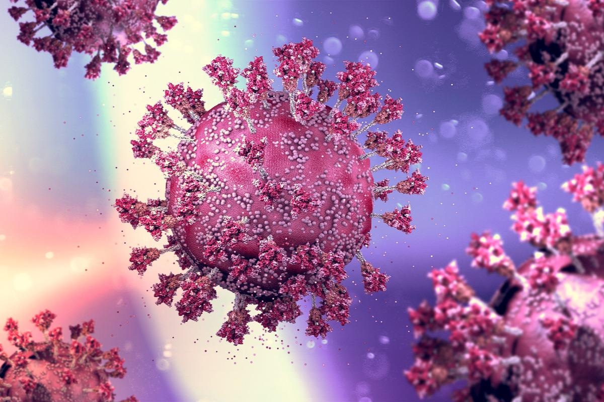
In a recent study posted to the bioRxiv* preprint server, researchers assessed the binding pocket of linoleic acid (LA), an essential free-fatty acid (FFA) in the receptor-binding domains (RBDs) of the spike (S) protein of beta-coronaviruses (b-CoVs).

Background
Human coronaviruses were previously only known to cause mild diseases of the upper respiratory tract until the emergence of the pathogenic coronaviruses and their mutant variants of concern (VOCs) which have substantially increased morbidity and mortality associated with b-CoVs infections.
The researchers of the present study had discovered LA bound to a hydrophobic pocket in the S RBD in their previous work. The FFA-binding was found to stabilize the locked conformation of the S protein and inhibit viral binding with the angiotensin-converting enzyme 2 (ACE2) receptors of the host cells, thereby preventing viral entry and replication in the host.
About the study
In the present study, researchers investigated if the LA-binding pocket is conserved across the RBDs of the S proteins of the following b-CoVs: severe acute respiratory syndrome CoV (SARS-CoV), SARS-CoV-2 and its VOCs (Alpha, Beta, Gamma, Delta, and Omicron), Middle East Respiratory Syndrome CoV (MERS-CoV), and the causative viruses of the common cold, namely, human CoV (HCoV)-HKU1 and HCoV-OC43.
Molecular dynamics (MD) simulations were performed to assess corroboration of the LA-Binding in the RBD pockets of the b-CoVs and SARS-CoV-2 VOCs. SARS-CoV S was analyzed by cryo-electron microscopy (cryo-EM), correlative light-electron microscopy (CLEM), and electron tomography of SARS-CoV-2-infected cells. LA-binding to S RBD was analyzed by surface plasmon resonance (SPR) experiments.
To assess the effects of LA-treatment on SARS-CoV-2-infected cells, Cancer coli-2 (Caco-2) cells overexpressing ACE2 (Caco-2-ACE2) were infected with a green fluorescent protein (GFP)-expressing SARS-CoV-2 and assessed at one-hour post-infection. Cell viability and viral replication were evaluated by fluorescence and brightfield microscopy at 35 hours post supplementation with LA.
Results and discussions
In this study, all the b-CoVs and SARS-CoV-2 VOCs were bound to LA, except HCoV-HKU1. However, one amino acid substitution of a residue that lines the entrance of the hydrophobic LA-binding pocket in the HCoV-HKU1 RBD was found to be adequate for restoring the LA-binding.
The cryo-EM, CLEM, and electron tomography analysis of SARS-CoV-2-infected cells showed that LA-treatment inhibited viral replication, and therefore lesser, deformed virions. LA-bound SARS-CoV S was found to adopt a hitherto elusive locked structure similar to LA-bound locked SARS-CoV-2 S, which is incompatible with ACE2 binding. Contrastingly, in the open conformation of SARS-CoV S, the hydrophobic pocket in the RBDs was devoid of LA.
In SPR experiments, LA bound with 72 nM and 96 nM binding affinities with the RBDs of SARS-CoV and MERS-CoV RBD, respectively. The binding affinity for SARS-CoV and SARS-CoV-2 were comparable. The RBDs of SARS-CoV-2 VOCs were found to bind with LA with binding affinities ranging from 50 nM to 87 nM, similar to SARS-CoV-2. This confirmed that the LA-binding pocket was conserved and was not affected by S mutations.
MD simulations showed three outcomes of (re)binding of LA with the RBD of SARS-CoV which were as follows: i) LA did not enter the hydrophobic pocket again during the one-ms simulation but showed close interaction with the receptor-binding motif (RBM) over a substantial period; ii) LA rebounded to the hydrophobic pocket post-removal, and iii) although LA entered the pocket, it could dissociate again.
In addition, the MD simulations showed that the binding of LA to the viral RBD is a dynamic process since the contacts formed between the lining residues of the pocket and LA varied with time and in-between experiments, indicative of various modes of binding. However, LA-binding to the entire S protein of SARS-CoV S showed that LA was bound in a stable manner to three pockets which were formed by adjacent RBDs within the S trimer protein with minimum molecular dynamics.
CLEM analysis of GFP-expressing cells showed a substantially greater number of virions in the untreated virus-infected cells (25 virions/micrograph) with smaller spherical virions compared to the LA treated cells post-infection (nine virions/micrograph) with larger irregularly shaped and highly elliptical and deformed virions. LA-treated cells also demonstrated lipid droplets within the cytoplasm (mainly in adipocytes) of cells that appeared dark on the micrographs, irrespective of whether they were infected or not. Additionally, substantial membrane remodeling was detected in the SARS-CoV-2-infected Caco-2-ACE2 cells.
The LA-mediated inhibition of viral replication probably occurred due to the inhibition of the cytoplasmic phospholipase A2 (cPLA2) enzyme via the formation of replication compartments within the cells. LA did not bind to HCoV-HKU1 S since this common cold-causing virus comprises a bulky glutamate E375 residue situated in front of the binding pocket that obstructs the entrance of the binding pocket.
Overall, the study findings showed that the LA-binding pocket is conserved across pathogenic b-CoVs except for HCoV-HKU1 and this pocket inhibits viral replication. LA treatment post-infection decreased the viral load in infected cells. This indicates that the LA-binding hydrophobic pocket could be a potential target of antiviral therapeutics.
*Important notice
bioRxiv publishes preliminary scientific reports that are not peer-reviewed and, therefore, should not be regarded as conclusive, guide clinical practice/health-related behavior, or treated as established information.
- Christine Toelzer, et al. (2022). The Free Fatty Acid-Binding Pocket is a Conserved Hallmark in Pathogenic b-Coronavirus Spike Proteins from SARS-CoV to Omicron. bioRxiv. doi: https://doi.org/10.1101/2022.04.22.489083 https://www.biorxiv.org/content/10.1101/2022.04.22.489083v1
Posted in: Medical Science News | Medical Research News | Disease/Infection News
Tags: ACE2, Adipocytes, Amino Acid, Angiotensin, Angiotensin-Converting Enzyme 2, binding affinity, Cancer, Cell, Cold, Common Cold, Coronavirus Disease COVID-19, Cytoplasm, Electron, Electron Microscopy, Elliptical, Enzyme, Fluorescence, Fluorescent Protein, Linoleic Acid, Membrane, MERS-CoV, Micrograph, Microscopy, Mortality, Omicron, Protein, Receptor, Respiratory, SARS, SARS-CoV-2, Severe Acute Respiratory, Severe Acute Respiratory Syndrome, Syndrome, Therapeutics, Tomography, Virus

Written by
Pooja Toshniwal Paharia
Dr. based clinical-radiological diagnosis and management of oral lesions and conditions and associated maxillofacial disorders.
Source: Read Full Article


