Skip to:
- What is Plasmonics?
- Applications
- Enhancing Raman Spectroscopy Signals
- Plasmonic Nanoscopy and Imaging
Plasma is one of the fundamental states of matter that is generated when a neutral gas is heated or subjected to a strong electromagnetic field, both of which can result in the breaking of bonds and the stripping of electrons from atomic nuclei.
Plasmonics is a field that is at the interface of optics and condensed matter, and it involves studying the phenomenon that is associated with plasmons: the oscillation of electrons in a plasma or surrounding the atomic nucleus of metals.
What is Plasmonics?
The resonance of surface plasmons occurs in metal nanoparticles when light leads to the oscillation of free electrons belonging to the nanoparticle. The incident light displaces the electrons with respect to the lattice ions. This results in the oscillations of the electron density. Upon excitation of the surface plasmon resonance, the electromagnetic fields are enhanced in the vicinity of the metal nanoparticles.
This enhanced field then generates thermal energy as the photons are converted into heat due to electron-electron scattering and electron-photon coupling. These nanoscale fields interact with cells, and both large and small biomolecules, both optically and thermally.
By controlling these interactions, it is possible to use this property for the sensing, analyzing, trapping, transporting, and manipulating of different cells and molecules. This has also led to the development of devices such as analytical tools for biology and nanomedicine. For example, metal nanoparticles are highly sensitive in detecting molecular-level biological processes.
Also, studies show that this technology can be used for plasmonic photothermal therapy for cancer.
Applications
Nanosensors
Detecting molecular interactions and dynamics at the smallest scales requires detecting molecules with high sensitivity. The resonance wavelength of a plasmon is sensitive to changes in refractive index that surround the nanoparticles due to the nanoscale localization and enhanced electromagnetic field. The intense and narrow plasmon resonance can be monitored to detect molecule-level changes in the local refractive index.
Using spectroscopic methods, plasmon resonance based sensors are being extensively used to detect molecules, biomolecules, and cellular signalling in vivo.
In a study, plasmon based nanosensors were used to measure the ligands that were derived from amyloid, measured at a concentration of as low as 100 fM.
One of the milestones for this process was to reach a single-molecule detection limit. This was achieved by increasing the sensitivity and plasmonic response of metal nanoparticles to these molecules by incorporating new structures, increasing the dielectric constant, etc.

Enhancing Raman Spectroscopy Signals
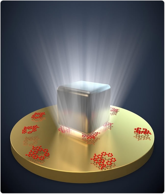
Raman spectroscopy is used to identify and detect molecules based on their specific vibrational energies. In this method, inelastically scattered electrons either lose or gain energy equal to their molecular vibrations. However, detecting small numbers of molecules is difficult through Raman scattering. The electromagnetic fields that are enhanced by plasmons can amplify Raman signals, leading to sensitive molecular detection.
Plasmonic Nanoscopy and Imaging
Optical imaging is often limited by the diffraction limit of light. Using near-field scanning microscopy and plasmonic nanoscopy, nanoscale biological medical imaging can be performed with a high signal-to-noise ratio, high spatial and temporal resolution, and low power of illumination.
This method has also been used to detect the fluorescence rate of single molecules based on their distance to laser-irradiated gold nanoparticles. This also functions as a nanoscopic probe.
'Chemical vision' is another newer application of plasmonic nanoscopy where the chemical structure of an object is identified with spatial resolution. For example, using this method, the surface chemical fingerprints of silicone were mapped in a study recently.
Sources
- https://www.ncbi.nlm.nih.gov/pmc/articles/PMC3991779/#R65
- https://iopscience.iop.org/article/10.1088/2040-8986/aaa114/pdf
- https://juniperpublishers.com/gjn/pdf/GJN.MS.ID.555646.pdf
Further Reading
- All Plasmonics Content
Last Updated: Aug 30, 2019

Written by
Dr. Surat P
Dr. Surat graduated with a Ph.D. in Cell Biology and Mechanobiology from the Tata Institute of Fundamental Research (Mumbai, India) in 2016. Prior to her Ph.D., Surat studied for a Bachelor of Science (B.Sc.) degree in Zoology, during which she was the recipient of anIndian Academy of SciencesSummer Fellowship to study the proteins involved in AIDs. She produces feature articles on a wide range of topics, such as medical ethics, data manipulation, pseudoscience and superstition, education, and human evolution. She is passionate about science communication and writes articles covering all areas of the life sciences.
Source: Read Full Article


