Despite the widespread use of medical-grade computed tomography (CT) in death investigations around the world, this imaging technique is often limited in its ability to accurately depict smaller anatomical structures or trauma at the microscopic level.
Recent investigations into the use of micro-CT (an imaging modality that is commonly used for industrial purposes) as a forensic imaging tool has demonstrated its usefulness as a complementary method to histology in post-mortem examinations.
Skip to:
- What is micro-CT?
- Micro-CT in forensics
- Validating micro-CT by comparison with histology
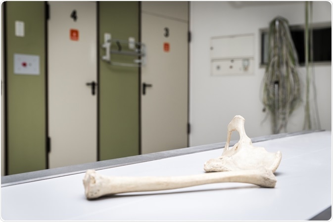 Miriam Doerr Martin Frommherz | Shutterstock
What is micro-CT?
When used within the industry, micro-CT is a useful tool for the non-destructive inspection of the internal surfaces and characteristics of small manufactured parts. This imaging technology is superior to the standard CT approach as it has a much higher resolution for visualizing minute details of samples. When applied in the industry, there is a reduced concern for exposing equipment to the high radiation levels of micro-CT as compared to a medical setting.
Micro-CT or microtomography, functions very similar to a CT machine commonly used in the medical setting; however, micro-CT produces images of significantly greater resolution of 100 nanometers (nm). This imaging technique can, therefore, provide high resolution three-dimensional (3D) images of material and biological samples by compiling a series of two-dimensional (2D) planar X-ray images.
Micro-CT in forensic science
In addition to high radiation doses, micro-CT is not commonly used in the clinical setting as it can create motion artifacts in images. Since these concerns are not applicable for analyzing cadaveric tissues in forensics, the use of post-mortem CT and more recently micro-CT has been used in medico-legal death investigations.
Micro-CT, in particular, has provided forensic scientists with information regarding specific injuries, such as pediatric rib fractures, in much greater detail compared to other imaging techniques.
Validating micro-CT by comparison with histology
A 2019 Forensic Science International paper aimed to validate the use of micro-CT in post-mortem studies by comparing the data obtained by this technology with histological analysis. In their study, three different specimens were used, including two adult laryngo-hyoid complexes obtained from suspected strangulation cases and a five-month old’s rib cage obtained from a suspected child abuse victim. Once the micro-CT images were obtained of these bones, the samples were submitted for histological analysis.
The study showed that micro-CT is a complementary method to histology, as the 3D model created by this imaging technique supported information from the analysis of the 2D histology slices. Out of the total of fourteen bone fractures that were identified in this study, micro-CT detected five in an initial scan and eight additional fractures in a second reading. In fact, micro-CT was found to identify additional features of trauma that were not originally detected in the histological analysis.
Since histology slices are taken at intervals of several millimeters (mm), the potential of missing small and subtle trauma markers is reduced when accompanied by micro-CT images. By using both histology and micro-CT together, this study found that the histopathologist could accurately determine whether a specific area of damage was real or an artifact from processing.
While micro-CT cannot replace the gold standard of histology altogether, it serves as a highly accurate wound analysis technique in forensics.
Sources
- Baier, W., Mangham, C., Warnett, J. M., Payne, M., Painter, M., & Williams, M. A. (2019). Using histology to evaluate micro-CT findings of trauma in three post-mortem samples – First steps towards method validation. Forensic Science International 297; 27-34. DOI: 10.1016/j.forsciint.2019.01.027.
- What is Micro-CT? An Introduction – Micro Photonics Inc.
Further Reading
- All Forensics Content
- Epigenetics and Forensics
- Chromatography in Forensic Science
- Overcoming DNA Degradation in Forensic Science
- Reducing PCR Inhibition in Forensic Science
Last Updated: May 29, 2019

Written by
Benedette Cuffari
After completing her Bachelor of Science in Toxicology with two minors in Spanish and Chemistry in 2016, Benedette continued her studies to complete her Master of Science in Toxicology in May of 2018.During graduate school, Benedette investigated the dermatotoxicity of mechlorethamine and bendamustine, which are two nitrogen mustard alkylating agents that are currently used in anticancer therapy.
Source: Read Full Article