
 insights from industryJason OtterstromBusiness Development Manager for EuropeIDEA Bio-Medical
In this interview, NewsMedical speaks with Dr. Jason Otterstrom about the benifits and challenges of using Zebrafish as an emerging model for life science reseach. We discuss how new automated approaches for analyzing micrscopy images of Zebrafish samples are enabling new insights, never before achievable. In particular, they focus on an automated, AI-powered analysis software, Athena. This innovation removes the bottleneck of manual quantification and is poised to enhance the rate of discovery to previously unachievable levels. Athena is now available for download in a novel pay-per-use basis.
Could you please provide us with an overview of why zebrafish are popular models for research purposes?
Zebrafish are a promising and exciting emergent model system for performing biomedical research in the life sciences. They have a number of advantages over existing mouse models, such as very fast reproductive times and a more economical husbandry. Additionally, higher experimental throughput is possible due to their short reproductive time.
Moreover, their genetic sequence has around 70% similarity to humans, which makes them genetically relevant, while also being amenable to genetic manipulation for introduction of fluorescent proteins as a reporter mechanism.
The size of the zebrafish is also an important advantage. Zebrafish form very small embryos and larvae, so in their young stages, they are transparent, making them very amenable to microscopy.
Even with bright-field imaging, the fish is clearly observable. Detailed internal anatomy can be seen along with biological functions happening in a living, multi-organ system, which is advantageous for pharmacokinetics, toxicity studies, and genetic knockouts.
There is also the benefit of the fluorescence giving a high contrast readout that is very binary. I believe there are many advantages that are going to push zebrafish into new fields in the coming decade.
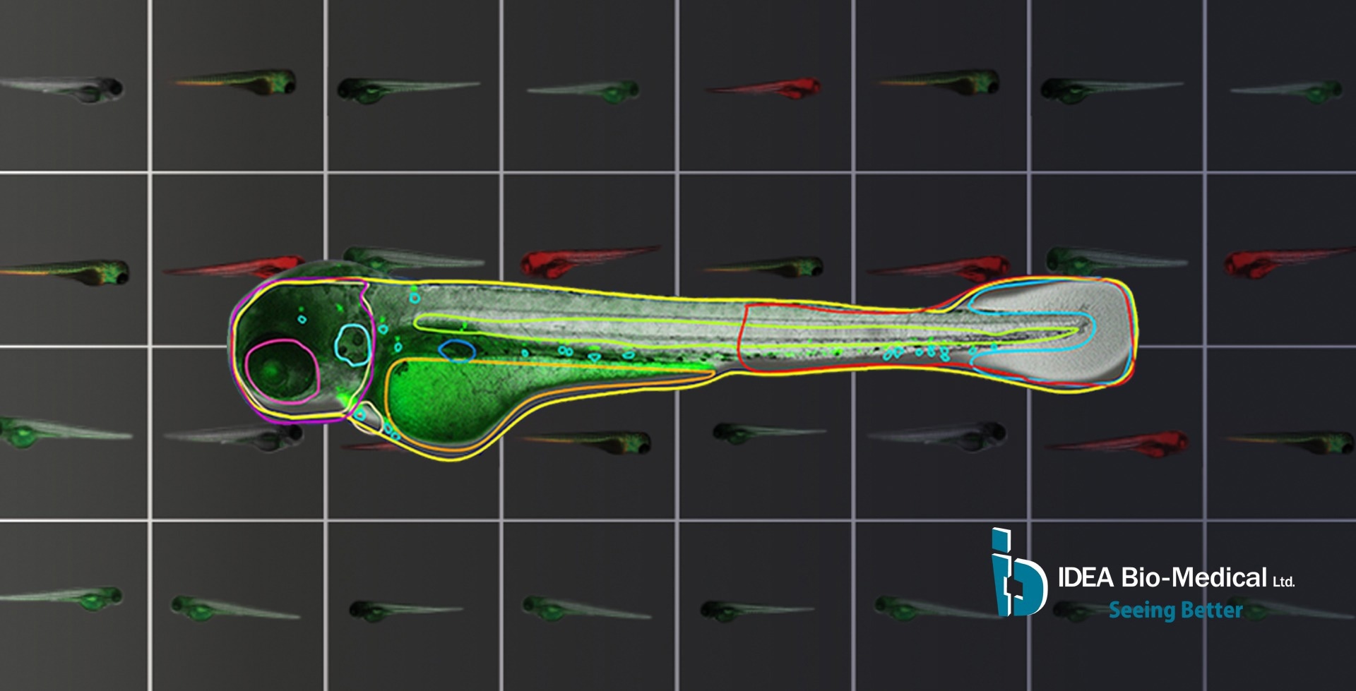
Image credit: IDEA Bio-Medical
What challenges do researchers face using zebrafish models?
With all the exciting potential that zebrafish hold for the future of life science research, there are several challenges when applying them as a model system for microscopy. While their transparency is an advantage and they can be imaged on many types of microscopes, placing them in the right orientation is a substantial challenge. This is because, depending on how they are oriented, you can see some of their internal anatomies better, or they can become obfuscated and unobservable, both in bright-field and in fluorescence.
Many researchers struggle with the challenge of getting the fish oriented to see the anatomy that is of interest with the highest fidelity.
There are different approaches to address this; some involve specialized alignment plates, others involve agarose gels – but none are fully meeting the throughput needs. Such approaches require a lot of manual intervention, contributing to the difficulty of overcoming this challenge.
Another challenge is, once the images have been acquired, to extract meaningful data from them. The images themselves to a human are easily interpretable; the fish and its organs can be seen, but a computer does not always register this.
Until now, many have relied on ImageJ and other types of programable software to manually identify areas of interest or simply look at the fluorescence channel and disregard anatomical context to obtain simplistic fluorescence readouts. As a result, meaningful information from disregarded anatomy is not considered.
Could you explain how IDEA Bio-Medical is able to offer potential solutions to these challenges?
IDEA Bio-Medical specializes in automating solutions for image acquisition and image analysis. Our Hermes platform, which includes the Hermes microscope and the accompanying Athena software, has been cited in over a hundred scientific journals supporting high impact research globally. As a small company, we work very closely with our clients and, through them, have recognized the need for image analysis in zebrafish.
At IDEA Bio-Medical, we come from a background of high-content screening where everything is automated. We have over a decade of experience now empowering life science researchers to automate their microscopy research in image acquisition and image analysis.

IDEA Bio-Medical at the annual Zebrafish Disease Models conference (ZDM). Image credit: IDEA Bio-Medical
Could you tell us the relationship that you have with your clients and how IDEA Bio-Medical helps support any changes in their research?
As a small company, we like to work very closely with our clients and through them, we came to realize the challenge zebrafish researchers face in their image analysis. Together with our clients, we have developed a novel, deep learning-based algorithm to identify the fish and its internal anatomy in bright-field images, a completely label-free image acquisition requirement.
From that image analysis, we are able to extract the fish contour and all of the internal anatomy. We then extract quantitative information about the morphology of the fish and its internal anatomy, using that information also to study the fluorescent signal that may be present. Studying the specific localization of the fluorescence within the zebrafish anatomy is possible as a result. Ultimately, we enable true high-content imaging in zebrafish for the first time.
How accessible is the Athena software to researchers as a whole, do they require exclusive access to the Hermes platform?
We are now making the software available to researchers who use any microscope, be it an upright or inverted stereo microscope, and no matter how many images they may need to analyze, be it a dozen or hundreds per week. This is something that we are very proud to be releasing to support the zebrafish community.
Up until now, this software package has only been part of our Hermes platform. Still, we are moving it to become a standalone product to support all zebrafish researchers. We are able to accept images from all types of microscopes, analyze them, and we have the capacity to accept image formats from all types of microscopes, be it open-source TIFF images or proprietary image formats.
The images can be acquired on upright microscopes, inverted microscopes, or stereo microscopes. In this way, we hope to support researchers who need to analyze a few dozen Zebrafish images per week or even hundreds per week. There is no throughput limit and by identifying all of this, we hope to empower zebrafish researchers to extract more from their data.
About Jason Otterstrom

Dr. Jason Otterstrom has a diverse background in biophysics including microscopy, optical design, image analysis and sample labeling. His expertise is adapting biological assays to benefit from utilization of automated microscopy methods. He obtained a Bachelor of Arts in Applied Physics at the University of Utah. After he obtained his PhD in Biophysics & Microscopy at Harvard University, He went on to obtain a Marie Curie fellowship to utilize super-resolution imaging at the Institute of Photonic Sciences (ICFO) near Barcelona, Spain.
As a business development manager for IDEA Bio-Medical , Jason supports clients with adapting their diverse experiments and assays to be performed in the context of automated microscopy on the company’s flagship product, the WiScan Hermes.
About IDEA Bio-Medical Ltd.
IDEA Bio-Medical is a company specializing in automated microscopy and image analysis for life science researchers. It was founded in 2007 through a partnership between YEDA (the Weizmann Institute’s commercialization arm) and IDEA Machine Development Design and Production Ltd (an innovation hub). IDEA’s products, the Hermes imaging system and Athena image analysis SW, have contributed to over 100 scientific publications in peer-reviewed magazines, globally supporting high impact science.
IDEA Bio-Medical currently focuses on empowering zebrafish researchers, specifically, to provide them with a reliable, robust solution for automated and unbiased Zebrafish image analysis by applying company’s knowledge and expertise.
To this end, IDEA developed a novel deep learning-based image analysis software for in-vivo zebrafish experiments. The software automatically detects zebrafish contour and their internal organs in brightfield with no required user input. The anatomy identified is coupled to fluorescence channels to permit anatomy-specific study of fluorescence changes. It is an affordable, user-friendly system designed specifically for reliable, automated zebrafish image-based analysis.
The software is available as a stand-alone product and accepts microscopy images in multiple image formats, including proprietary ones. It is suited for researchers who only image and analyze a handful of fish per week, as well as researchers imaging hundreds and thousands of fish in multi-well plates for large scale screens. IDEA Bio-Medical is offering a novel pay-per-use model to access the software to enable flexible access. So, all researchers using manual microscopes or automated systems from other vendors, can readily use IDEA’s Zfish software to extract quantitative, meaningful information from their Zebrafish images when they need it.
Reach out to IDEA Bio-Medical on our website's contact form, and more information can be found on the zebrafish analysis software product page.
Sponsored Content Policy: News-Medical.net publishes articles and related content that may be derived from sources where we have existing commercial relationships, provided such content adds value to the core editorial ethos of News-Medical.Net which is to educate and inform site visitors interested in medical research, science, medical devices and treatments.


