
Metastasis accounts for approximately 90 percent of mortality in breast cancer patients. During the last few decades, there has been significant progress in understanding genetic, molecular and signaling mechanisms underpinning cancer cell migration.
Biologists from Lawrence Livermore National Laboratory (LLNL) found another mechanism that affects the maintenance and expansion of malignant cells: electric signals in the tumor microenvironment.
All cells can generate bioelectric signals through their plasma membrane, and therefore naturally exist in our bodies. Cancer growth interferes with local ionic, membrane and epithelium environments, resulting in small electrical changes in the tumor microenvironment.
Electrical fields (EFs) have significant effects on cancer cell migration, but their role as a regulator of cancer progression and metastasis is poorly understood.
Despite the implementation of advanced detection technologies, the prevalence of metastatic breast cancer at initial diagnosis has remained stagnant in the United States since 1975.
The LLNL-University of California, Davis collaborative team used unique probe systems to characterize the electrical properties of cancer cells and how they migrate under an EF. Earlier studies showed that bioelectric characteristics of cancer tissue differs from normal tissue and may change during cancer development.
“Our data contribute to an improved understanding of breast cancer metastasis, providing new evidence in support of an electrical mechanism that contributes to this phenomenon,” said LLNL biologist Gaby Loots, co-lead author of a paper appearing in Scientific Reports.
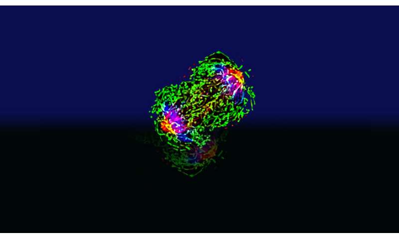
In the research, subcutaneous tumors were established from a triple-negative murine breast cancer cell line (4T1) and electric currents and potential areas where tumors could develop were measured using vibrating probes and glass microelectrodes, respectively. Steady outward and inward currents could be detected at different positions on the tumor surface, and magnitudes of the electric currents on the tumor surface strongly correlated with tumor weights.
Potential measurements also showed the non-homogeneous intra-tumor electric potentials. Cancer cell migration was then surveyed in the presence of EFs in vitro. Parental 4T1 cells and metastatic sub-lines in isolation showed random migration in EFs of physiological strength, whereas cells in monolayer migrated collectively to the anode.
“Cancer was long considered a disease defined and driven by genetic evolution, which was mapped to the signaling pathways that regulate cell growth or motility, but our research shows that there is increasing evidence that the maintenance and expansion of malignant cells also strongly depend on external signals from the tumor microenvironment,” Loots said.
Due to differences in metabolism and segregation of ions, local electrical properties changed and induced small direct current electrical fields naturally in live tissues. It has been shown to closely associate with cancer growth and other biological processes, such as wound healing. For instance, electric currents/fields at wounds are readily measurable and can persist from hours to weeks. Similarly, cancer growth interferes with local ionic, membrane and transepithelial (membranous tissue composed of one or more layers of cells that form the covering of most internal and external surfaces of the body and its organs) environments, and therefore produce local electric fields.
The team’s results revealed that the tumor did generate an electric field at the tumor surface, and tumor electric fields increase with the size of tumors.
Source: Read Full Article


